Heart Drawing Biology
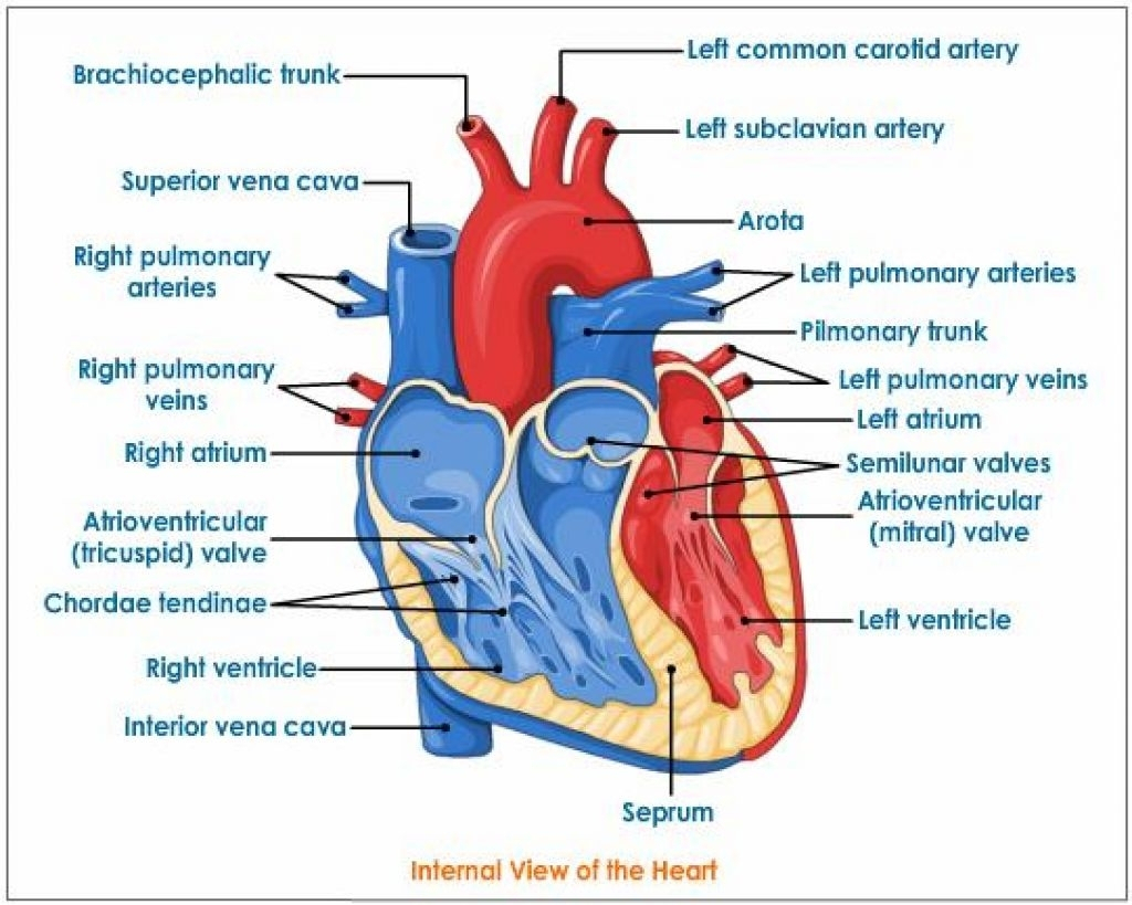
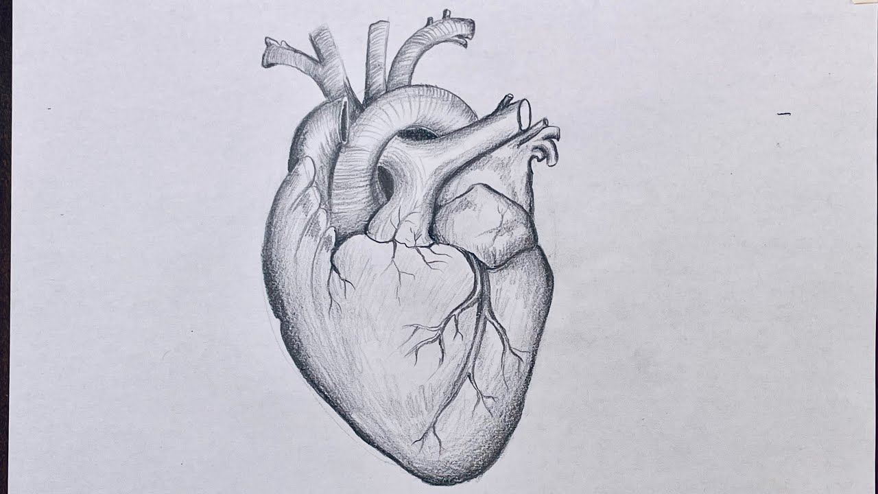
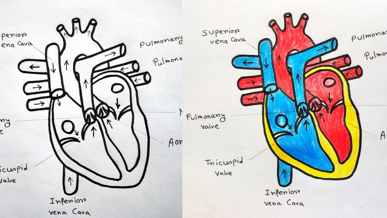
Heart Drawing Biology - The atria are the two superior chambers of the heart and the ventricles are the two inferior chambers of the heart. Then, fill in the base of the heart with the right and left ventricles and the right and left atriums. On its superior end, the base of the heart is attached to the aorta, pulmonary arteries and veins, and the vena cava. Drag and drop the text labels onto the boxes next to the diagram. The heart is made up of four chambers: Plus, you may just learn something new along the way. Web the heart is located in the thoracic cavity medial to the lungs and posterior to the sternum. The oxygenated blood is then pumped to the rest of the body. Sketch out a basic outline of the heart, using our tutorial as a guide. The upper two chambers of the heart are called auricles. Sketch out a basic outline of the heart, using our tutorial as a guide. The heart wall is made up of three layers: Web in this interactive, you can label parts of the human heart. Students who are unwilling to handle hearts, but keen to see what is going on could take digital photographs of the heart in. Web your heart is located in the front of your chest. The heart is divided into four chambers: In this lecture, dr mike shows the two best ways to draw and label the heart! Web how to draw human heart step by step, easy trick to draw human heart, how to draw the diagram of human heart, easily drawing diagram of human heart for class 10 student.more. Rishi is a pediatric infectious disease physician and works at khan academy. It includes a worksheet with the basic information they need to produce a biological drawing similar to what they should be producing in their pags. Web your heart is located in the front of your chest. Web 48k views 1 year ago cardiovascular system. In this video i will be. The heart is made up of four chambers: On its superior end, the base of the heart is attached to the aorta, pulmonary arteries and veins, and the vena cava. How to draw human heart easily step by step. Web to draw an anatomical heart realistically, pay attention to the proportions and positioning of the different parts of the heart, as well as their texture and color. Web drawing a human heart is easier than you may think. The heart is divided into four chambers: Web prepare a handout with. The atria are the two superior chambers of the heart and the ventricles are the two inferior chambers of the heart. Web how to draw heart | biology drawing for science students. We will then proceed to shape the heart, slowly refining it with our pencils into a. Web the heart is a muscular organ that pumps blood around the. The inferior tip of the heart, known as the apex, rests just superior to the diaphragm. Then, fill in the base of the heart with the right and left ventricles and the right and left atriums. Rishi is a pediatric infectious disease physician and works at khan academy. Web how to draw human heart step by step, easy trick to. The aim of these posts is to explicitly. Web to draw the internal structure of the heart, start by sketching the 2 pulmonary veins to the lower left of the aorta and the bottom of the inferior vena cava slightly to the right of that. Web the heart is located in the thoracic cavity medial to the lungs and posterior. This worksheet is designed to help a level students perfect their biological drawing technique. Web drawing a human heart is easier than you may think. The lower two chambers of the heart are called ventricles. The atria are the two superior chambers of the heart and the ventricles are the two inferior chambers of the heart. It sits slightly behind. Students who are unwilling to handle hearts, but keen to see what is going on could take digital photographs of the heart in. On its superior end, the base of the heart is attached to the aorta, pulmonary arteries and veins, and the vena cava. Web your heart is located in the front of your chest. Web learn how blood. The atria are the two superior chambers of the heart and the ventricles are the two inferior chambers of the heart. This video will help you to draw internal structure of human heart ( ಮಾನವ ಹೃದಯ ) in simple way. Web this post will focus on how i teach the structure of the heart so pupils can identify the four. In this lecture, dr mike shows the two best ways to draw and label the heart! Web the heart is a muscular organ that pumps blood around the body by circulating it through the circulatory/vascular system. The upper two chambers of the heart are called auricles. Web 48k views 1 year ago cardiovascular system. In this video i will be. Sketch out a basic outline of the heart, using our tutorial as a guide. Then, fill in the base of the heart with the right and left ventricles and the right and left atriums. Web internal structures of the heart. Web to draw an anatomical heart realistically, pay attention to the proportions and positioning of the different parts of the. It includes a worksheet with the basic information they need to produce a biological drawing similar to what they should be producing in their pags. Web the heart is located in the thoracic cavity medial to the lungs and posterior to the sternum. Web internal structures of the heart. Rishi is a pediatric infectious disease physician and works at khan academy. Web how to draw heart | biology drawing for science students. Web in this interactive, you can label parts of the human heart. The inferior tip of the heart, known as the apex, rests just superior to the diaphragm. Web the heart is a muscular organ that pumps blood around the body by circulating it through the circulatory/vascular system. This video will help you to draw internal structure of human heart ( ಮಾನವ ಹೃದಯ ) in simple way. Plus, you may just learn something new along the way. We will then proceed to shape the heart, slowly refining it with our pencils into a. This worksheet is designed to help a level students perfect their biological drawing technique. Then, fill in the base of the heart with the right and left ventricles and the right and left atriums. Right atrium, right ventricle, left atrium, and left ventricle. The aim of these posts is to explicitly. Your ribcage protects your heart, everyone’s heart is a slightly different.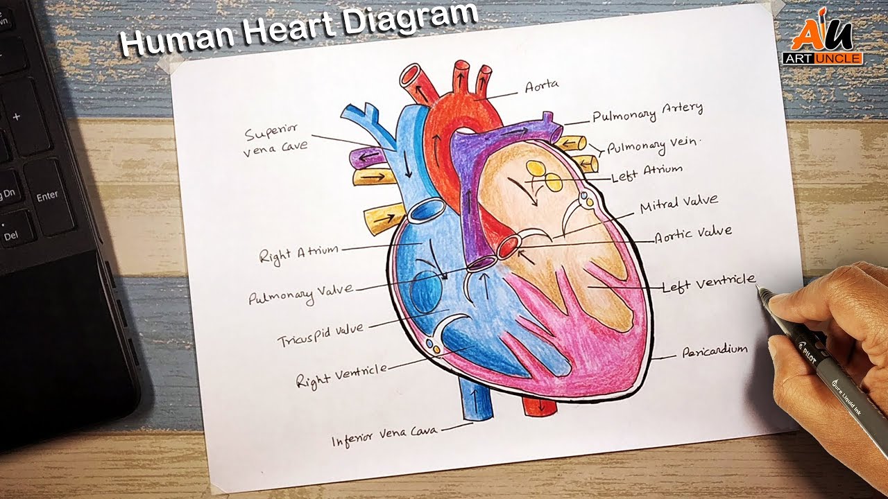
How to draw human heart_diagram Life processes NCERT class 10

PAG 2.1 The Heart biological Drawing OCR A Teaching Resources

how to draw human heart diagram easy/human heart drawing YouTube
Heart And Labels Drawing at GetDrawings Free download

How to Draw the Internal Structure of the Heart (with Pictures)
How to draw Heart Biology drawing for science students YouTube

Healthcare and Medical Education Drawing Chart of Human Heart Anatomy

Cardiac cycle and the Human Heart A* understanding for iGCSE Biology 2
How to draw Human Heart with colour Human Heart labelled diagram

How To Draw Human Heart Diagram
Selecting Or Hovering Over A Box Will Highlight Each Area In The Diagram.
Web This Is A Quick Way To Learn How To Draw The Heart And Some Of The Associated Structures
The Oxygenated Blood Is Then Pumped To The Rest Of The Body.
Web Learn How Blood Flows Through The Heart, And Understand The Difference Between Systemic And Pulmonary Blood Flow.
Related Post: