Heart Drawing Anatomy
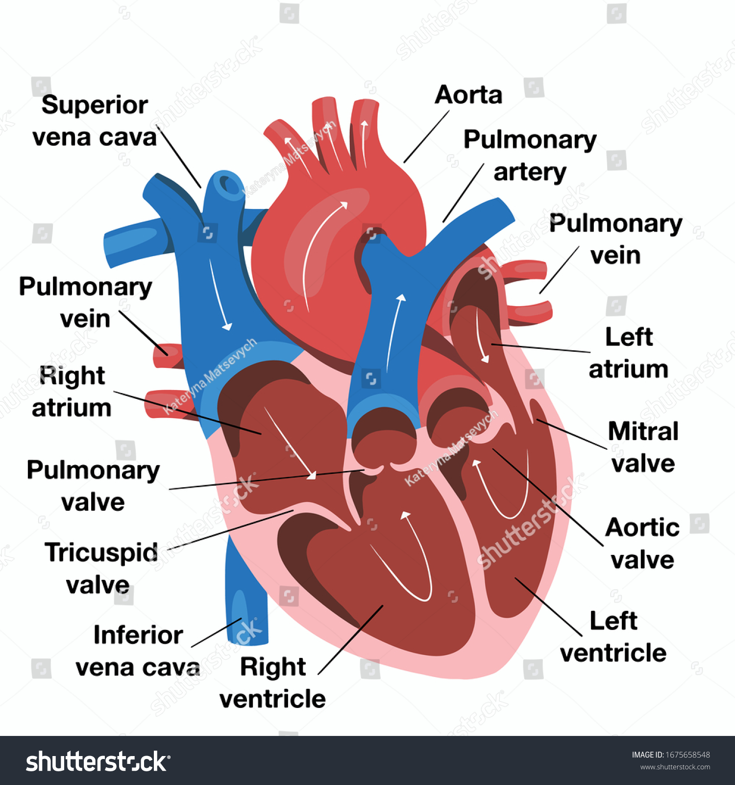
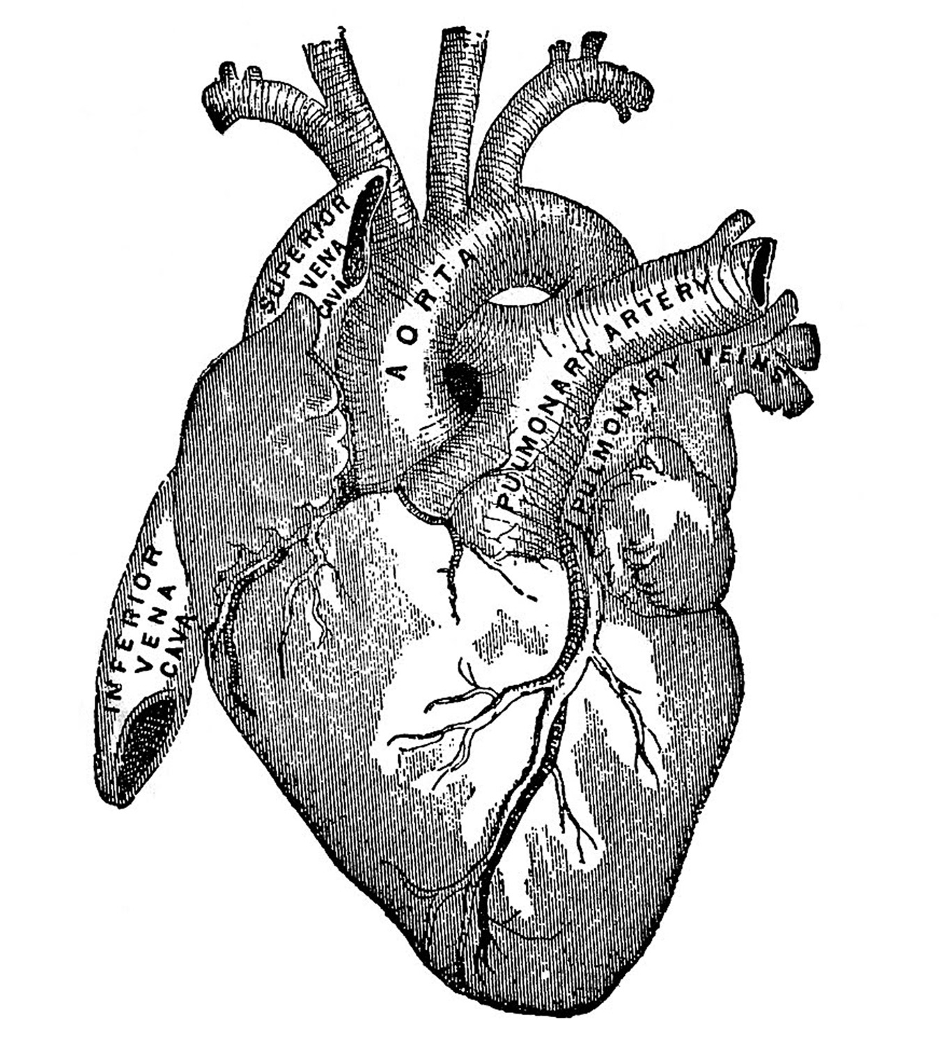
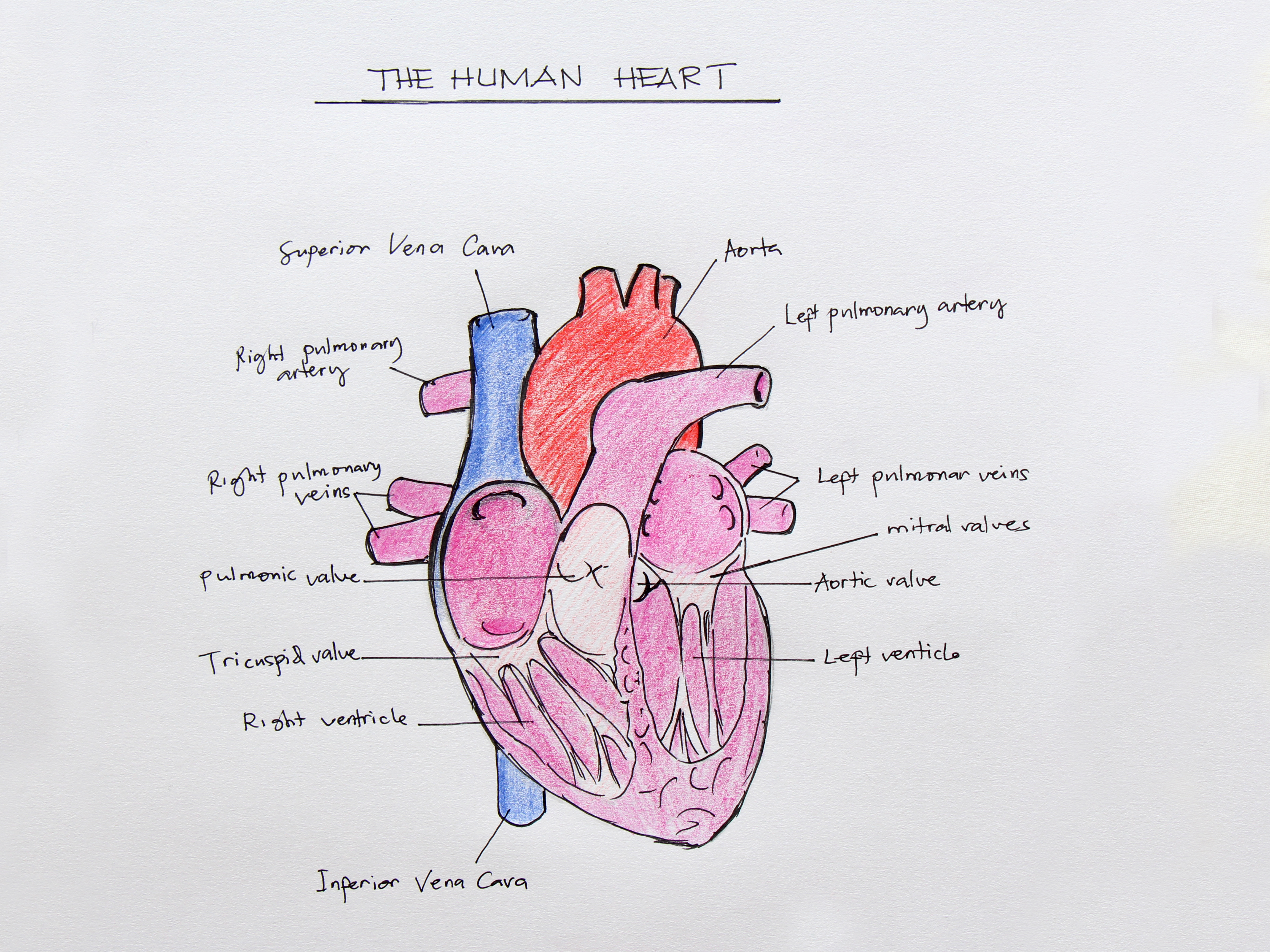
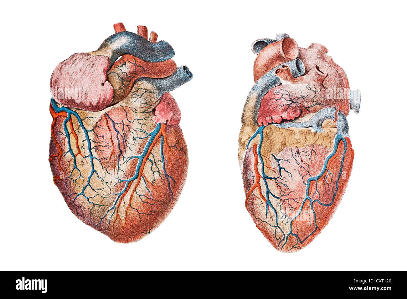
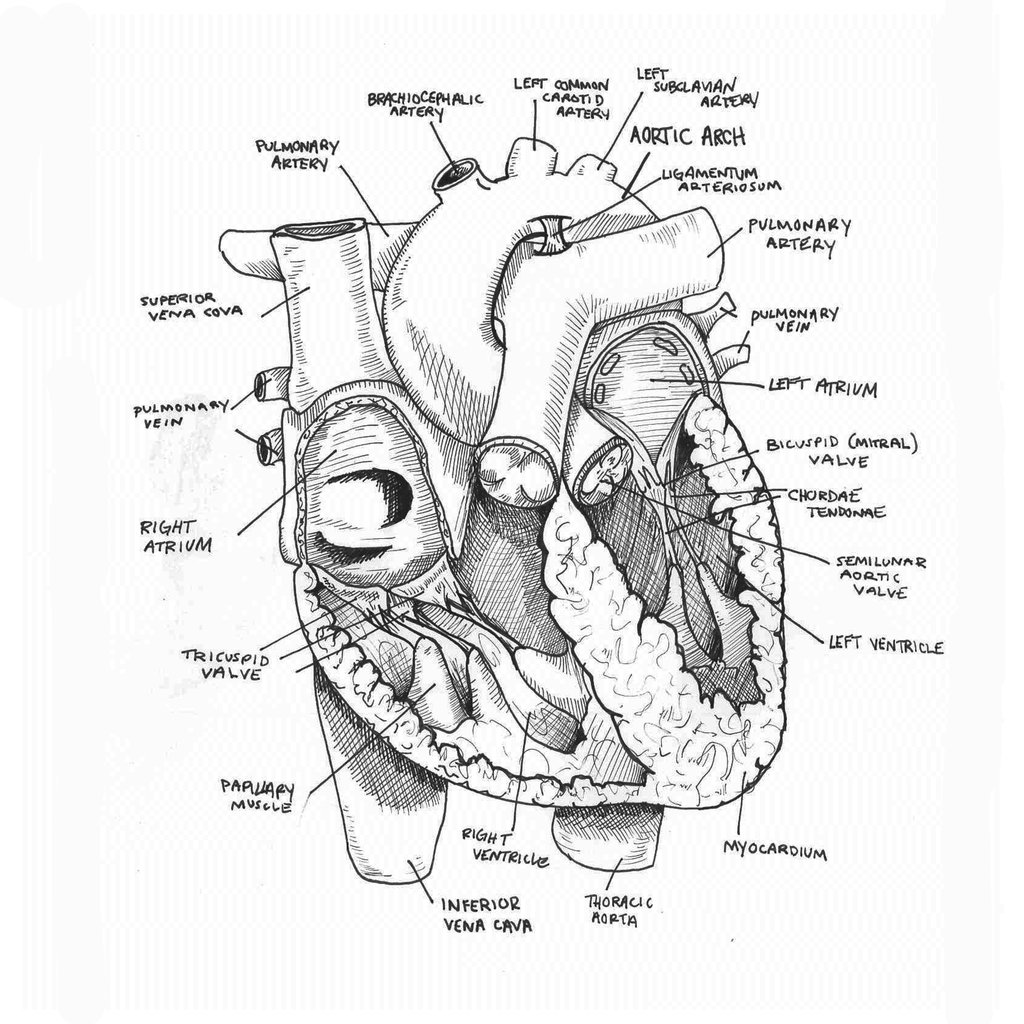
Heart Drawing Anatomy - For now, we shall be drawing the outline of the base of the heart, and this is the area that houses the ventricles. Less searching, more finding with getty images. Get free printable coloring page of this drawing. The right margin is the small section of the right atrium that extends between the superior and inferior vena cava. The inferior tip of the heart, known as the apex, rests just superior to the diaphragm. Web these anatomical heart medical illustrations are highly detailed drawings that blend art with science. Web dr matt & dr mike. Plus, you may just learn something new along the way. In this lecture, dr mike shows the two best ways to draw. Web the intricate anatomy of the heart can be challenging to grasp, and so i hope you find this tool to be helpful in visualizing the cardiac system. From the openstax anatomy and physiology book. Web study the anatomy of the heart, so you know where each part is located and how it interacts with other parts. Take the time to practice drawing each part individually before sketching the entire heart. Draw the human heart shape. There is also a murmurs tab that includes some of the more common murmurs we come across. Right, left, superior, and inferior: Web to draw the internal structure of the heart, start by sketching the 2 pulmonary veins to the lower left of the aorta and the bottom of the inferior vena cava slightly to the right of that. The user can show or hide the anatomical labels which provide a useful tool to create illustrations perfectly adapted for teaching. The right margin is the small section of the right atrium that extends between the superior and inferior vena cava. Get free printable coloring page of this drawing. Explore the beauty of anatomical heart drawings and unleash your creativity. Web learn how to draw a human heart with these 15 easy human heart drawing ideas with step by step sketch outline, printables and coloring pages. In this lecture, dr mike shows the two best ways to draw. Base (posterior), diaphragmatic (inferior), sternocostal (anterior), and left and right pulmonary. Use some curved lines for this aorta with a small oval shape at the tip of it. Draw the human heart shape. Web learn how to draw a human heart with these 15 easy human heart drawing ideas with step by step sketch outline, printables and coloring pages. Web in animals with lungs —amphibians, reptiles, birds, and mammals—the heart shows. Get free printable coloring page of this drawing. Web your heart sure does work hard, but that doesn’t mean you have to work hard to draw it! Sketch out a basic outline of the heart, using our tutorial as a guide. There is also a murmurs tab that includes some of the more common murmurs we come across. Included below. Web these anatomical heart medical illustrations are highly detailed drawings that blend art with science. Get free printable coloring page of this drawing. Web your heart sure does work hard, but that doesn’t mean you have to work hard to draw it! Explore the beauty of anatomical heart drawings and unleash your creativity. Outline a simple heart shape. There are lots of small parts that we will be drawing throughout this guide on how to draw a realistic heart, so for this first step we will be keeping things a bit simple. Web dr matt & dr mike. Included below are a magnificent color heart illustration, along with four monotype prints, which are possibly woodcuts, engravings, or lithographs.. Web the heart is located in the thoracic cavity medial to the lungs and posterior to the sternum. Web the intricate anatomy of the heart can be challenging to grasp, and so i hope you find this tool to be helpful in visualizing the cardiac system. Web this interactive atlas of human heart anatomy is based on medical illustrations and. Web learn how to draw a human heart with these 15 easy human heart drawing ideas with step by step sketch outline, printables and coloring pages. 48k views 1 year ago cardiovascular system. It’s an example of a drawing that can give you a basic idea of what a human heart sketch should be look like. There is also a. Less searching, more finding with getty images. Drawing a human heart is easier than you may think. Then, fill in the base of the heart with the right and left ventricles and the right and left atriums. There are lots of small parts that we will be drawing throughout this guide on how to draw a realistic heart, so for. Then, fill in the base of the heart with the right and left ventricles and the right and left atriums. There are lots of small parts that we will be drawing throughout this guide on how to draw a realistic heart, so for this first step we will be keeping things a bit simple. From the openstax anatomy and physiology. Web your heart sure does work hard, but that doesn’t mean you have to work hard to draw it! The average human heart weighs. Sketch out a basic outline of the heart, using our tutorial as a guide. Discover top ideas to create stunning and realistic anatomical heart artwork that will leave a lasting impression. 48k views 1 year ago. Web to draw an anatomical heart realistically, pay attention to the proportions and positioning of the different parts of the heart, as well as their texture and color. It’s an example of a drawing that can give you a basic idea of what a human heart sketch should be look like. Base (posterior), diaphragmatic (inferior), sternocostal (anterior), and left and right pulmonary surfaces. The heart has five surfaces: Two atria and two ventricles. Sketch out a basic outline of the heart, using our tutorial as a guide. Web the heart is located in the thoracic cavity medial to the lungs and posterior to the sternum. From the openstax anatomy and physiology book. Plus, you may just learn something new along the way. Use some curved lines for this aorta with a small oval shape at the tip of it. Web your heart sure does work hard, but that doesn’t mean you have to work hard to draw it! The inferior tip of the heart, known as the apex, rests just superior to the diaphragm. By following the simple steps, you too can easily draw a perfect human heart. 48k views 1 year ago cardiovascular system. The user can show or hide the anatomical labels which provide a useful tool to create illustrations perfectly adapted for teaching. On its superior end, the base of the heart is attached to the aorta, pulmonary arteries and veins, and the vena cava.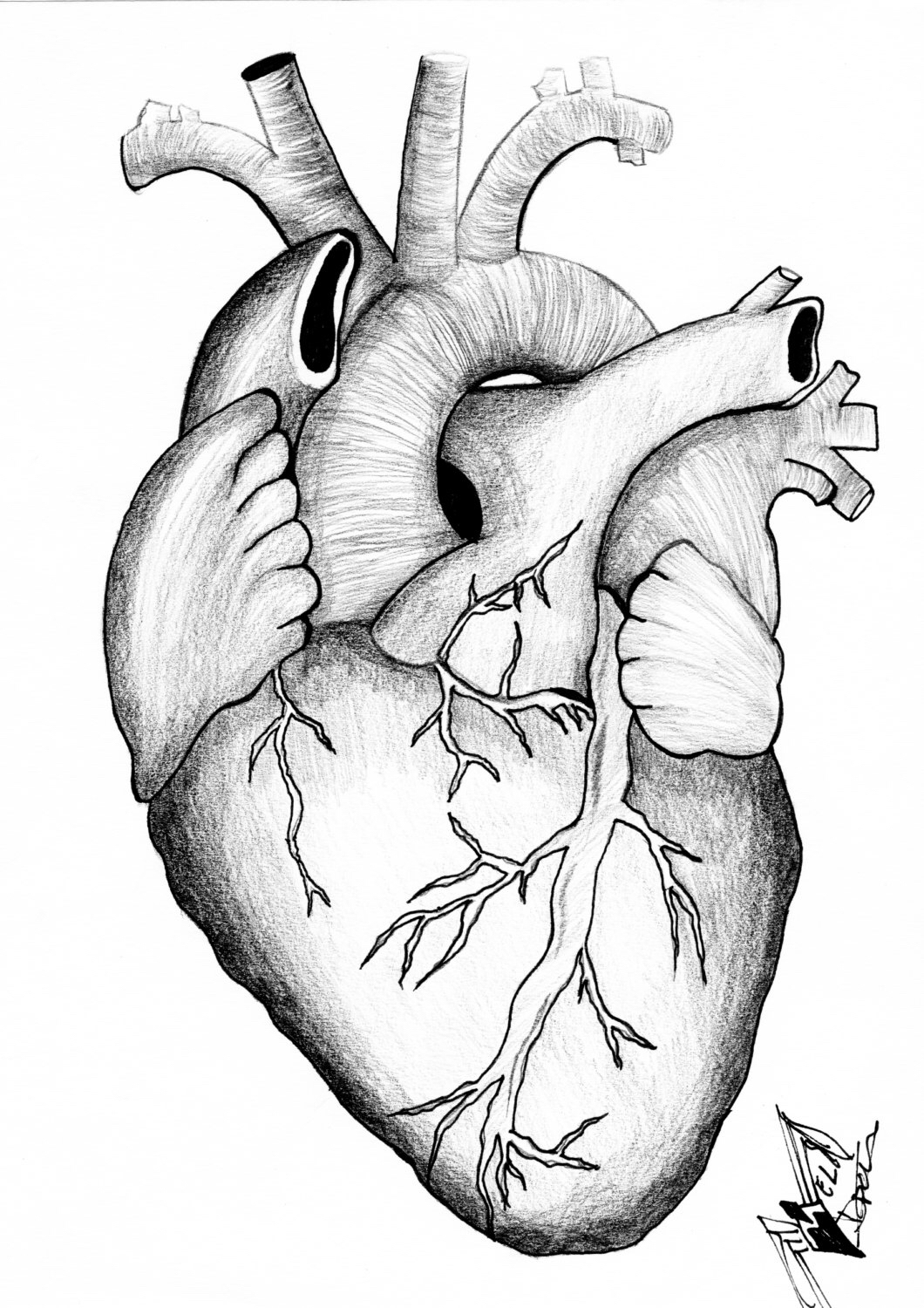
Anatomical Heart Drawing at GetDrawings Free download
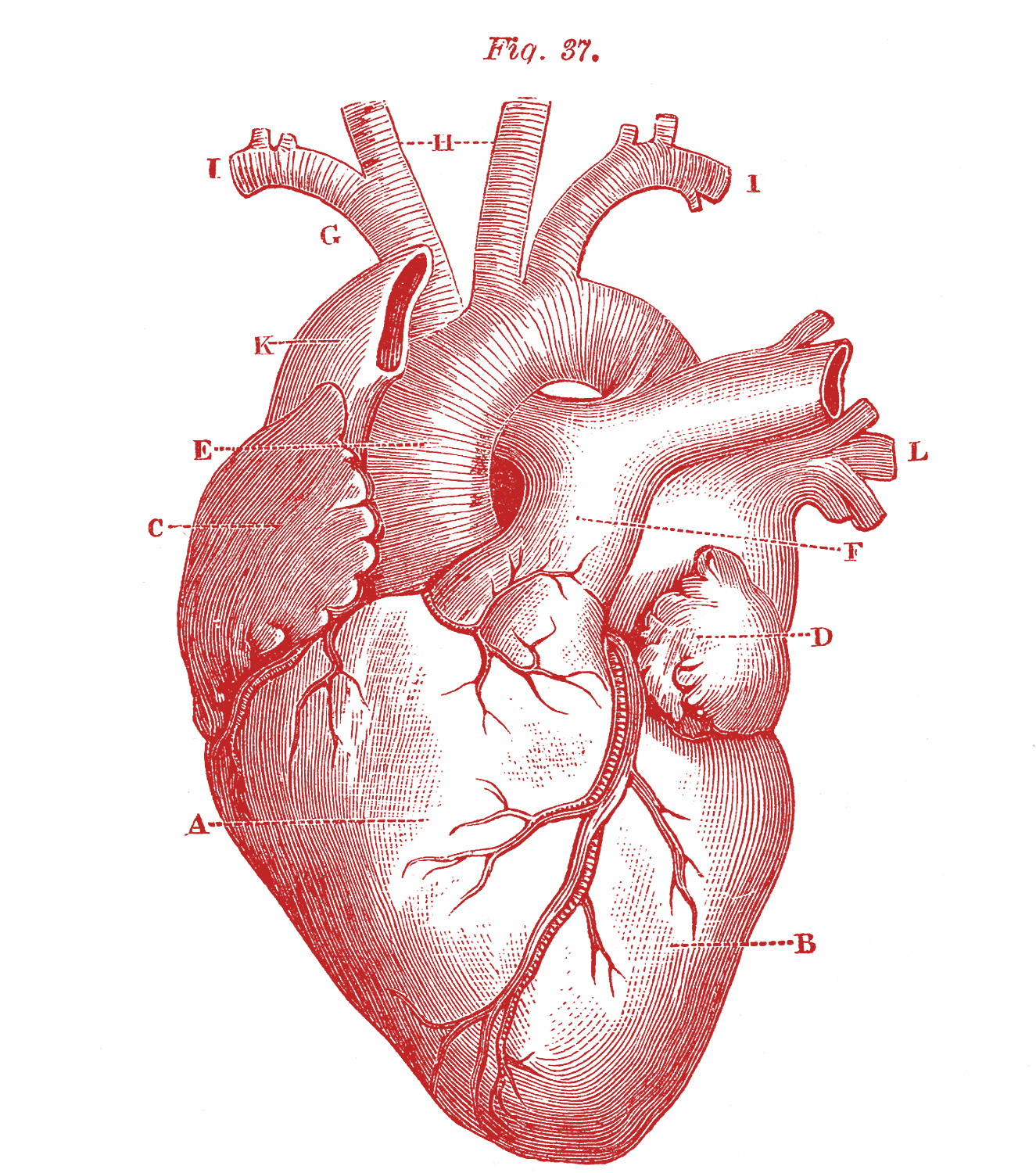
9 Anatomical Heart Drawings! The Graphics Fairy

How to Draw a Human Heart 11 Steps (with Pictures) wikiHow
Hand Drawn Illustration Human Heart Anatomy Stock Vector (Royalty Free
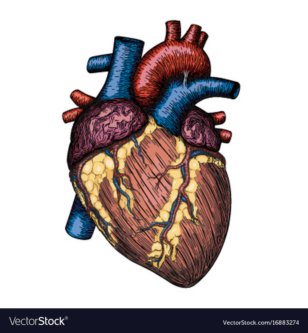
Human heart hand drawn anatomical sketch Vector Image
9 Anatomical Heart Drawings! The Graphics Fairy
Anatomical Drawing Heart at GetDrawings Free download
Anatomical heart drawing hires stock photography and images Alamy

External Structure Of Heart Anatomy Diagram
Anatomical Drawing Heart at GetDrawings Free download
Less Searching, More Finding With Getty Images.
Included Below Are A Magnificent Color Heart Illustration, Along With Four Monotype Prints, Which Are Possibly Woodcuts, Engravings, Or Lithographs.
Web The Intricate Anatomy Of The Heart Can Be Challenging To Grasp, And So I Hope You Find This Tool To Be Helpful In Visualizing The Cardiac System.
For Now, We Shall Be Drawing The Outline Of The Base Of The Heart, And This Is The Area That Houses The Ventricles.
Related Post: