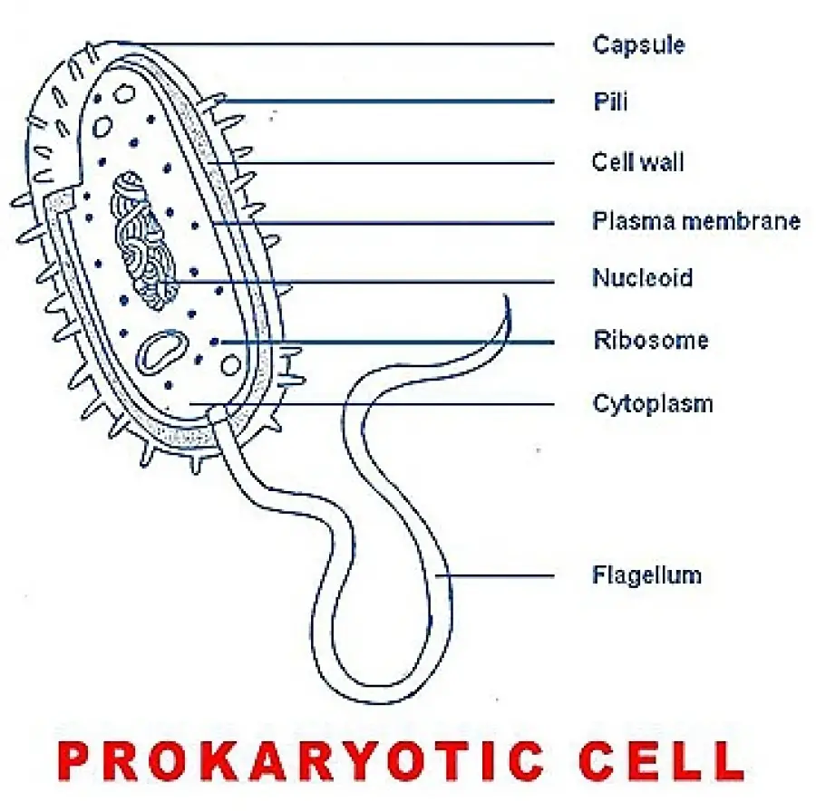
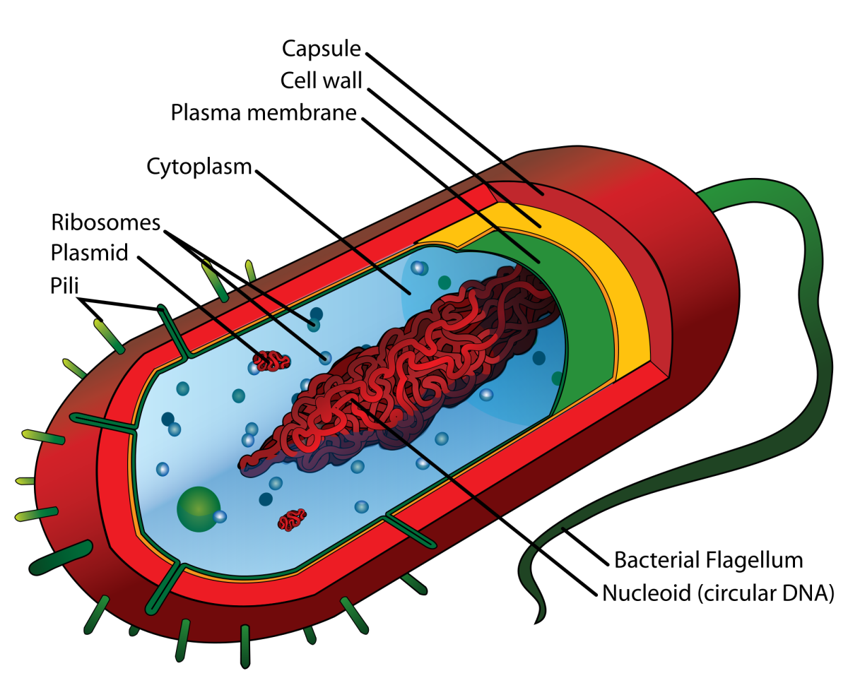
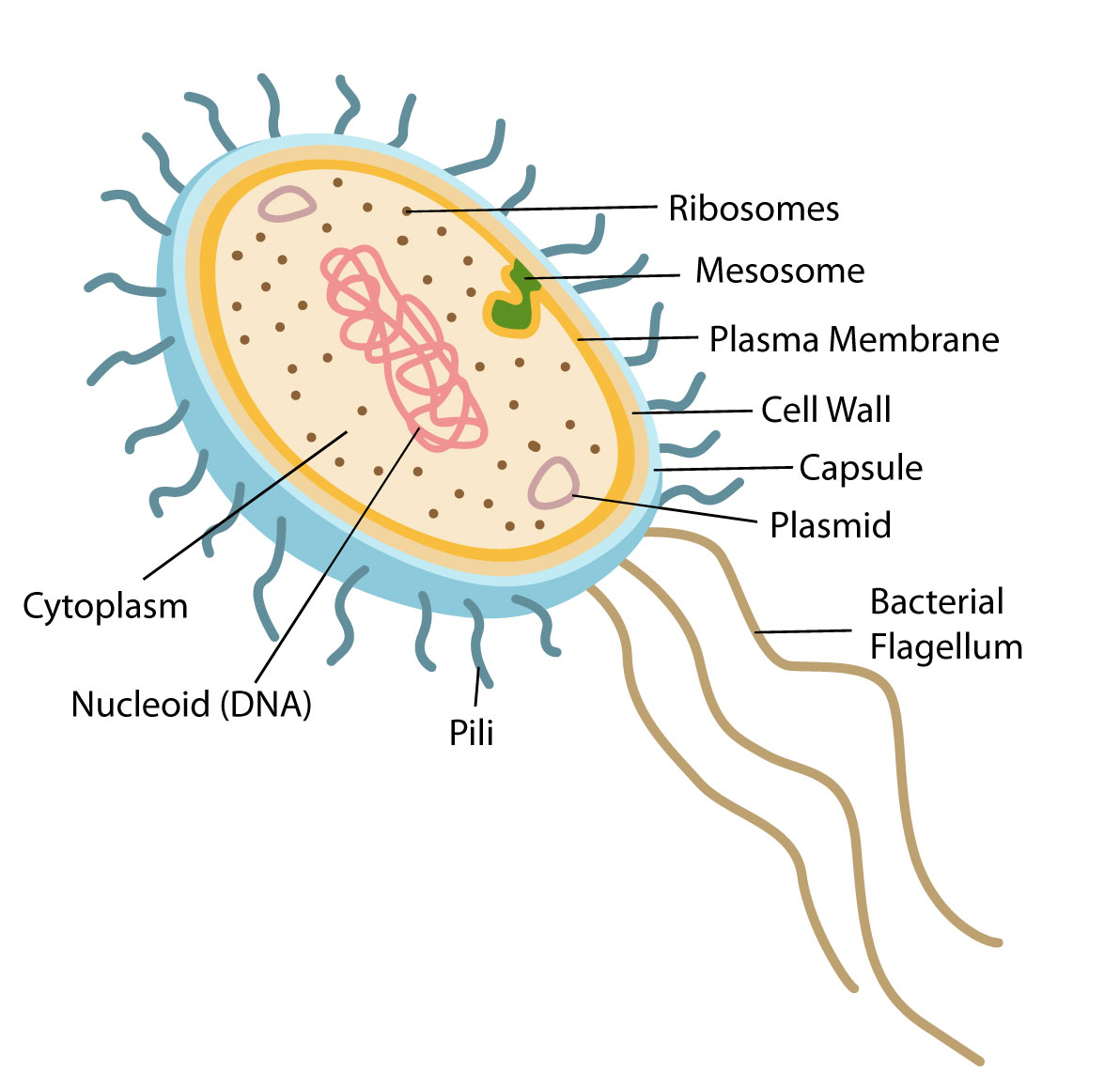
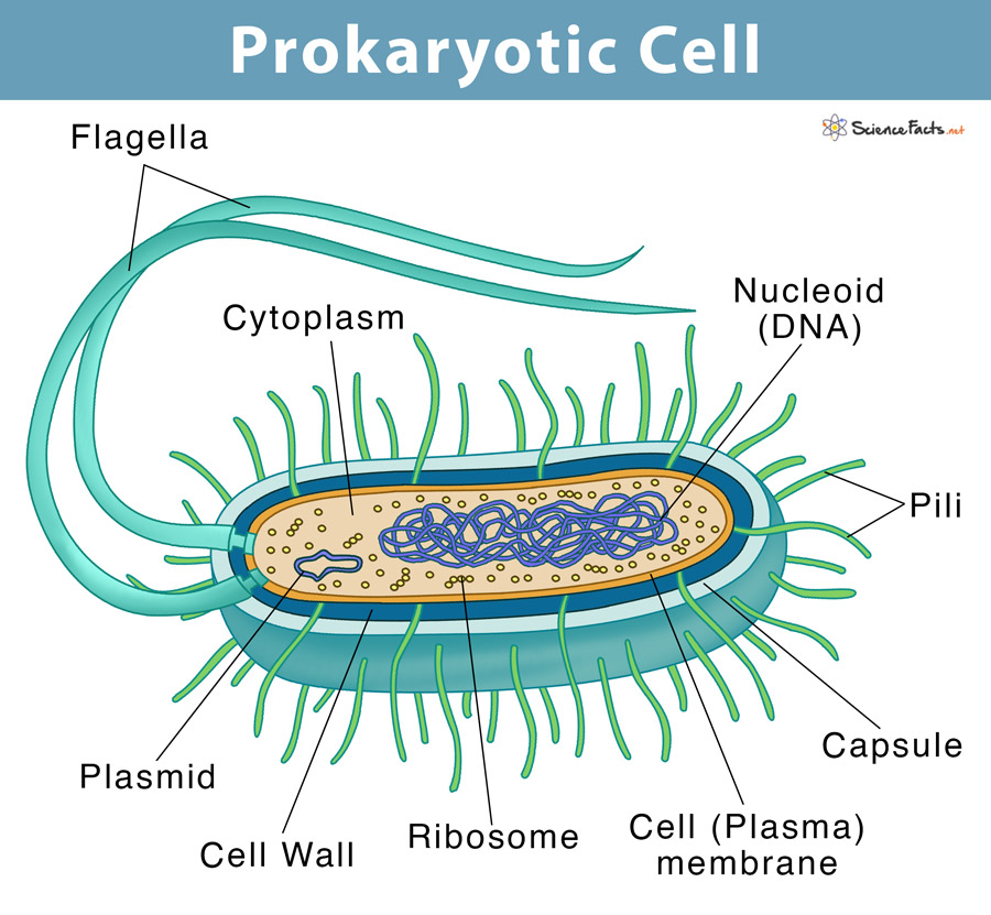
Drawing Of A Prokaryotic Cell
Drawing Of A Prokaryotic Cell - The plasma membrane, or cell membrane, is the phospholipid layer that surrounds the cell and protects it from the outside environment. The figure below shows the sizes of prokaryotic, bacterial, and eukaryotic, plant and animal, cells as well as other molecules and organisms on a logarithmic. Web the nucleus (plural, nuclei) houses the cell’s genetic material, or dna, and is also the site of synthesis for ribosomes, the cellular machines that assemble proteins. Web step by step and simple way to draw prokaryotic cell with easy methods. Web identify the three most common shapes of prokaryotic cells. A darkened region called the nucleoid (figure 2). Both prokaryotic and eukaryotic cells have structures in common. Web figure 4.5 this figure shows the generalized structure of a prokaryotic cell. Describe a typical prokaryotic cell. Web unit 1 intro to biology. Web identify the three most common shapes of prokaryotic cells. As i go, i give tips on drawing the various structures and how to label them. These cells are structurally simpler and smaller than their eukaryotic counterparts, the cells that make up fungi, plants, and animals. Web distinguish between prokaryotic cells and eukaryotic cells in terms of structure, size, and the types of organisms that have these cell types. Web prokaryotic cell diagram drawing easy and step by step / how to draw and label prokaryotic cell in this video, i will learn how to draw and label a prokaryot. Prokaryotic dna is found in the central part of the cell: Web identify the three most common shapes of prokaryotic cells. Unit 6 structure of a cell. This video explains how to draw prokaryotic cell in easy. Web unlike eukaryotic cells, prokaryotic cells lack a true nucleus and complex organelles, but they have structures such as a cell wall, capsule, cytoplasm, and flagella that support their functions and survival. Some prokaryotes have flagella, pili, or. Web the nucleus (plural, nuclei) houses the cell’s genetic material, or dna, and is also the site of synthesis for ribosomes, the cellular machines that assemble proteins. The features of a typical prokaryotic cell are shown. Web typical prokaryotic cells range from 0.1 to 5.0 micrometers (μm) in diameter and are significantly smaller than. The features of a typical prokaryotic cell are shown. Web identify the three most common shapes of prokaryotic cells. Diagram of a typical prokaryotic cell. Unit 7 more about cells. The figure below shows the sizes of prokaryotic, bacterial, and eukaryotic, plant and animal, cells as well as other molecules and organisms on a logarithmic. The other structures shown are present in some, but not all, bacteria. The figure below shows the sizes of prokaryotic, bacterial, and eukaryotic, plant and animal, cells as well as other molecules and organisms on a logarithmic. Web unit 1 intro to biology. What are the roles of flagella and endospores in prokaryotes? Unit 6 structure of a cell. Describe a typical prokaryotic cell. All cells have a plasma membrane, ribosomes, cytoplasm, and dna. Unit 6 structure of a cell. Web unlike eukaryotic cells, prokaryotic cells lack a true nucleus and complex organelles, but they have structures such as a cell wall, capsule, cytoplasm, and flagella that support their functions and survival. Prokaryotic dna is found in the central. Unit 8 membranes and transport. Web figure 4.5 this figure shows the generalized structure of a prokaryotic cell. These neat, well labelled and colorful diagrams will make your answers look more. Some prokaryotes may have additional structures such as a capsule, flagella, and pili. The figure below shows the sizes of prokaryotic, bacterial, and eukaryotic, plant and animal, cells as. Unit 8 membranes and transport. Web many prokaryotic cells have sphere, rod, or spiral shapes (as shown below). The plasma membrane, or cell membrane, is the phospholipid layer that surrounds the cell and protects it from the outside environment. Web i draw a bacterial cell to show you how to make an accurate biological drawing of a prokaryotic cell. Web. The composition of the cell wall differs significantly between the domains bacteria and archaea, the two. Web identify the three most common shapes of prokaryotic cells. As i go, i give tips on drawing the various structures and how to label them. What are the roles of flagella and endospores in prokaryotes? Diagram of a typical prokaryotic cell. Unit 4 elements of life. Both prokaryotic and eukaryotic cells have structures in common. Web the nucleus (plural, nuclei) houses the cell’s genetic material, or dna, and is also the site of synthesis for ribosomes, the cellular machines that assemble proteins. Web what is a prokaryotic cell. How it looks like under a microscope. Web distinguish between prokaryotic cells and eukaryotic cells in terms of structure, size, and the types of organisms that have these cell types. Web identify the three most common shapes of prokaryotic cells. Web i draw a bacterial cell to show you how to make an accurate biological drawing of a prokaryotic cell. All prokaryotes have chromosomal dna localized in. As i go, i give tips on drawing the various structures and how to label them. Web typical prokaryotic cells range from 0.1 to 5.0 micrometers (μm) in diameter and are significantly smaller than eukaryotic cells, which usually have diameters ranging from 10 to 100 μm. The photosynthetic prokaryotes include cyanobacteria that perform photosynthesis. Both prokaryotic and eukaryotic cells have. Prokaryotes include bacteria and archaea. Prokaryotic dna is found in the central part of the cell: What are the roles of flagella and endospores in prokaryotes? This video explains how to draw prokaryotic cell in easy. The composition of the cell wall differs significantly between the domains bacteria and archaea, the two. Web i am demonstrating the colorful diagram of prokaryotic cells step by step which you can draw very easily. Both prokaryotic and eukaryotic cells have structures in common. The plasma membrane, or cell membrane, is the phospholipid layer that surrounds the cell and protects it from the outside environment. Some prokaryotes have flagella, pili, or. Learn its structure, with diagram, facts, characteristics, parts, functions, & cell division. Web unit 1 intro to biology. These neat, well labelled and colorful diagrams will make your answers look more. Bacteria and archaea differ in the lipid composition of their cell membranes and the characteristics of the cell wall. Describe a typical prokaryotic cell. Web identify the three most common shapes of prokaryotic cells. The photosynthetic prokaryotes include cyanobacteria that perform photosynthesis.
Draw a well labelled diagram of a prokaryotic cell.
Simple Prokaryotic Cell Diagram
Prokaryotic Cell Structure A Visual Guide Owlcation
Prokaryotic Cell Structure, Characteristics & Function

Prokaryotic cell structure diagram, Stock vector Colourbox
Prokaryotic Cell Definition, Examples, & Structure
:max_bytes(150000):strip_icc()/bacteria_cell_drawing-5786db0a5f9b5831b54f017c.jpg)
Prokaryotic Cells Structure, Function, and Definition

Draw A Well Labelled Diagram Of Typical Prokaryotic Cell The Best

What's In A Cell? Library For Kids

Cell Biology Glossary Cell Architecture of Prokaryotes (Bacteria
In The Following Sections, We’ll Walk Through The Structure Of A Prokaryotic Cell, Starting On The Outside And Moving Towards The Inside Of The Cell.
The Other Structures Shown Are Present In Some, But Not All, Bacteria.
How It Looks Like Under A Microscope.
Web Many Prokaryotic Cells Have Sphere, Rod, Or Spiral Shapes (As Shown Below).
Related Post: